Hypertrophic cardiomyopathy (HCM) is a myocardial disorder defined by left ventricular (LV) hypertrophy in the absence of other causes1-3

  • Hypercontractility and subsequent hypertrophy of the ventricular walls are characteristics of HCM1-3
  • The thickening of the LV wall cannot be explained by another cardiac or systemic disease2-4
  • This progressive and adverse cardiac debilitation can occur over many years, leading to chronic and acute health complications that may often be irreversible1,2

One U.S. study estimated that at least 85% of Americans with HCM are likely undiagnosed1,3,5


Normal
heart

Heart with
HCM

HCM is a lifelong, chronic condition that can worsen over time, resulting in serious and ongoing health issues1,2

HCM is the most common genetic cardiac disorder1-3

  • In the majority of cases, HCM is inherited with a ~50% risk of transmission to offspring1-3
  • Sarcomere mutations are the most common genetic basis of HCM2,3
    • The sarcomere is the basic contractile unit of striated muscle6
    • Sarcomeres are responsible for contraction and relaxation of all heart muscle cells6
Learn about the pathophysiology of HCM

Nonobstructive vs. obstructive HCM: What to look for

  • Nonobstructive HCM
    • The LV muscle is thickened to ≥15 mm, but there is little to no blockage of blood flow out of the LV1-3*
    • The peak resting and stress gradient (LV outflow tract [LVOT]) is <30 mmHg2,3
  • Obstructive HCM
    • The blockage caused by the thickened heart muscle, referred to as LVOT obstruction, reduces the blood flow out of the left ventricle1-3
    • The peak resting and stress LVOT gradients are ≥30 mmHg1-3

~70-75%


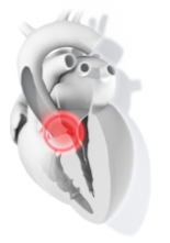
of patients with HCM have LVOT obstruction, either at rest or with provocation1,3

Nonobstructive
HCM

Obstructive
HCM

    Both forms can lead to adverse complications and outcomes; however, the risk of heart failure is higher in patients with obstructive HCM1,2

HCM is a chronic, progressive disease with poorer outcomes when it is associated with LVOT obstruction1-3

Footnote

* The clinical criterion for diagnosing HCM for a patient who has a family history of HCM or a positive genetic test is ≥13 mm LV wall thickness.2

HCM=Hypertropic cardiomyopathy; LV=Left ventricular; LVOT=Left ventricular outflow tract.

References

  1. Naidu, Srihari S, editor. Hypertrophic Cardiomyopathy. Second ed., Springer International Publishing, 2019.
  2. Gersh BJ, et al. 2011 ACCF/AHA guideline for the diagnosis and treatment of hypertrophic cardiomyopathy. J Thorac Cardiovasc Surg. 2011;142(6):e153-203.
  3. Ommen SR, et al. 2020 AHA/ACC guideline for the diagnosis and treatment of patients with hypertrophic cardiomyopathy: a report of the American College of Cardiology/American Heart Association Joint Committee on Clinical Practice Guidelines. Circulation. 2020;142(25):e558-e631.
  4. Nagueh SF, et al. Tissue Doppler imaging consistently detects myocardial abnormalities in patients with hypertrophic cardiomyopathy and provides a novel means for an early diagnosis before and independently of hypertrophy. Circulation. 2001 Jul 10;104(2):128-30.
  5. Maron MS, et al. Occurrence of clinically diagnosed hypertrophic cardiomyopathy in the United States. Am J Cardiol. 2016;117(10):1651-4.
  6. Garfinkel AC, et al. Genetic pathogenesis of hypertrophic and dilated cardiomyopathy. Heart Fail Clin. 2018;14(2):139-46.